Example
Developing a radiologic interpretation system of malignant bone tumors by artificial intelligence
Social background
Although a primary malignant bone tumor is a so-called orphan cancer, many of the patients develop it in the childhood and adolescent and young adult (AYA) generations, and the Ministry of Health, Labour and Welfare has recently spoken of the need for medical care and assistance measures against this disease. One of the main prognostic factors is whether there is any metastasis at initial diagnosis, and thus how to reduce oversight at initial diagnosis and delay in diagnosis are the most important considerations. However, due to its rarity, there are few expert physicians and thus there is no end to the number of patients who have died after the spread of the cancer due to delays in diagnosis, highlighting the need to improve the system of diagnosis.
Objectives of activities
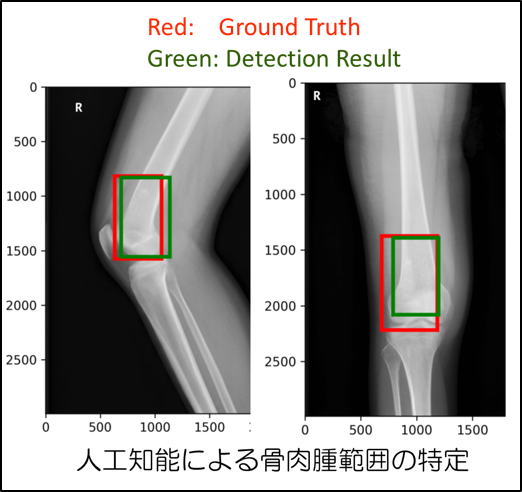
Recently, the development of artificial intelligence (AI) has been remarkable, and its application to the field of medicine has been expected. Studies on imaging and pathological diagnostic technologies using AI are currently being conducted throughout the world; however, artificial intelligence technology for bones are still developing, not to mention its application to the treatment of bone tumors. In this significantly important, challenging study, we aim to develop an imaging diagnostic support technology for primary malignant bone tumors, thereby reducing the need for oversight at the first radiographic (Xp) inspection on outpatients, in particular, to save as many children’s lives as possible.
Overview of activities
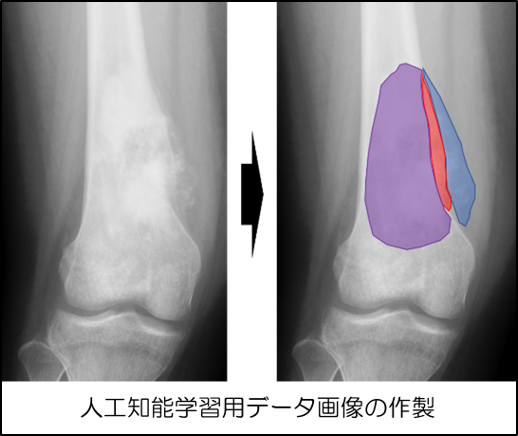
We are provided with data from bone and soft tissue sarcoma diagnosis facilities, centered on Okayama University Hospital and promoting the development of artificial intelligence. Furthermore, by collaborating with external companies dealing with artificial intelligence, we aim to incorporate artificial intelligence into a diagnosis system.
Expected effects
It is expected that this study will enable all clinical sites to secure a constant level of interpretation of radiograms.
Representative